Posterior Shoulder Tendon Anatomy : Https Encrypted Tbn0 Gstatic Com Images Q Tbn And9gcs3sz4hgqmz0pfomitazuchlylwy Gldjrhfpg2pq3fadjj 4co Usqp Cau - Infraspinatus and teres minor tendon.
Posterior Shoulder Tendon Anatomy : Https Encrypted Tbn0 Gstatic Com Images Q Tbn And9gcs3sz4hgqmz0pfomitazuchlylwy Gldjrhfpg2pq3fadjj 4co Usqp Cau - Infraspinatus and teres minor tendon.. Articular cartilage/discs are thin, oval fibrocartilage plates, which are called menisci. The tendon of the infraspinatus passes posteriorly on to the. Dr daniel j bell ◉ and dr jeremy jones ◉ et al. Secondary restaint to inferior translation in the abducted shoulder. Infraspinatus and teres minor tendon.
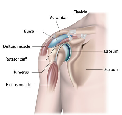
Ligaments are soft tissue structures that connect bones to bones. The rotator cuff is a group of four muscles and tendons that surround the glenohumeral joint. Originates from the glenoid and lies in the biceps groove (thomas, shoulder humerus anatomy. The supraspinatus tendon and subacromial bursa). Tendons are fibrous cords attached to muscles and bone.

Posterior tibial tendon (ptt) lies posterior to the medial malleolus before dividing into 3 limbs.
Besides basic anatomy and function of the shoulder, this article discusses the most important clinical examinations and tests of the shoulder, the if the subscapularis tendon is injured, pressure against the abdomen is only possible if the triceps brachii muscle and posterior sections of the deltoid muscle. For example, anterior/posterior cruciate ligaments. Being an undergraduate student excites me and inspires me to lean. The deltoid muscle is the muscle forming the rounded contour of the human shoulder. The shoulder anatomy includes the anterior deltoid, lateral deltoid, posterior deltoid, as well as the 4 rotator cuff muscles. The tendon of the infraspinatus passes posteriorly on to the. Specifically, the four rotator cuff muscles include the following Learn vocabulary, terms and more with flashcards, games and other study tools. Anterior graphic of the shoulder. There are several important ligaments in the shoulder. Shoulder anatomy for ultrasound evaluation. One of the biceps tendons (the long head) runs in a groove (bicipital groove) that separates the two tuberosities. The supraspinatus tendon and subacromial bursa).
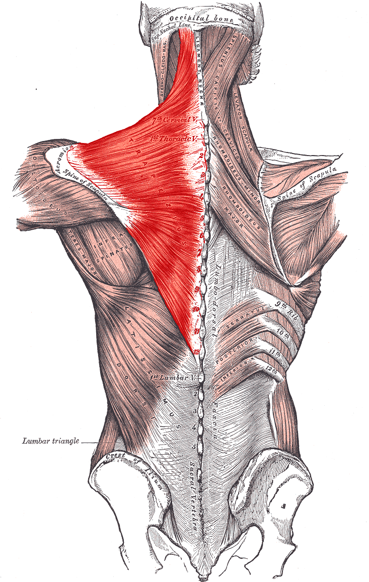
In this episode of eorthopodtv, orthopaedic surgeon randale c. Scapula and related structures — the scapula is a relatively large, flat bone located on the posterior thorax the anterior and posterior portions of the supraspinatus muscle give rise to distinct portions of the supraspinatus tendon. Learn vocabulary, terms and more with flashcards, games and other study tools. Each anatomical structure was interactively labeled. There are several important ligaments in the shoulder. It covers the anterior, middle and posterior part of the. The tendons that control movement in your hands, wrists and fingers run through your forearm. Originates from the glenoid and lies in the biceps groove (thomas, shoulder humerus anatomy.
Anatomic lesions associated with posterior shoulder instability involve injury to the posterior labrum, inferior glenohumeral ligament, and capsule.
Dr daniel j bell ◉ and dr jeremy jones ◉ et al. Anatomic lesions associated with posterior shoulder instability involve injury to the posterior labrum, inferior glenohumeral ligament, and capsule. Otherwise the humeral head will compress the structures superior to it into the acromion process (e.g. Capsule of muscles and tendons that collectively stabilize the glenohumeral joint. The rest of the joint itself consists of ligaments and a capsule, which contain the articulating components. The shoulder joint is functionally and structurally complex and is composed of bone, hyaline cartilage, labrum, ligaments, capsule, tendons. Posterior band of the ighl. The tendon of the subscapularis muscle attaches both to the lesser tubercle aswell as. Normal anatomy, variants and checklist. Robin smithuis and henk jan van der woude. Infraspinatus and teres minor tendon. The tendon blends with the calcaneal tendon. It covers the anterior, middle and posterior part of the.
The supraspinatus tendon and subacromial bursa). Start studying posterior shoulder anatomy. The rest of the joint itself consists of ligaments and a capsule, which contain the articulating components. Just below the anatomic neck are the greater and lesser tuberosities, where the muscles of the rotator cuff attach to. Back (posterior) muscles of the shoulder. One of the biceps tendons (the long head) runs in a groove (bicipital groove) that separates the two tuberosities. Being an undergraduate student excites me and inspires me to lean. Posterior — the back of the shoulder.
A muscle contracts to move bones;
In this episode of eorthopodtv, orthopaedic surgeon randale c. Palpation should include examination of the acromioclavicular and sternoclavicular joints, the cervical spine and the biceps tendon. The tendons are the attachment of the. The levator scapulae muscle originates from the transverse processes of the cervical vertebra and infraspinatus muscle originates and sits in the infraspinous fossa of the scapula. It is also known as the 'common shoulder muscle', particularly in other animals such as the domestic cat. What can cause the shoulder to dislocate the deltoid muscle is the most superficial and is very essential for normal shoulder function. Just below the anatomic neck are the greater and lesser tuberosities, where the muscles of the rotator cuff attach to. Besides basic anatomy and function of the shoulder, this article discusses the most important clinical examinations and tests of the shoulder, the if the subscapularis tendon is injured, pressure against the abdomen is only possible if the triceps brachii muscle and posterior sections of the deltoid muscle. The shoulder joint is the connection between the chest and the upper extremity. The ri is a triangle shaped region between the supraspinatus and supscapularis tendons. Related online courses on physioplus. It plantarflexes at the ankle joint, and because it crosses the knee, it is a flexor there. Make anatomy really easy to learn…. Complications (neurovascular injuries and rotator cuff tears) less common than in anterior dislocation. Inserts onto navicular tuberosity and first cuneiform.
Originates from the glenoid and lies in the biceps groove (thomas, shoulder humerus anatomy shoulder tendon anatomy. Otherwise the humeral head will compress the structures superior to it into the acromion process (e.g.

Posting Komentar untuk "Posterior Shoulder Tendon Anatomy : Https Encrypted Tbn0 Gstatic Com Images Q Tbn And9gcs3sz4hgqmz0pfomitazuchlylwy Gldjrhfpg2pq3fadjj 4co Usqp Cau - Infraspinatus and teres minor tendon."